Featured
- Get link
- X
- Other Apps
Types of Medical Imaging: Technologies along with Career Options

Imaging technologists who are interested by pursuing one-of-a-kind varieties of scientific imaging modalities, enhancing their competencies and understanding, and advancing their careers, ought to bear in mind a bachelor’s in imaging sciences read more:- serverpress
Types of Medical Imaging Technologies
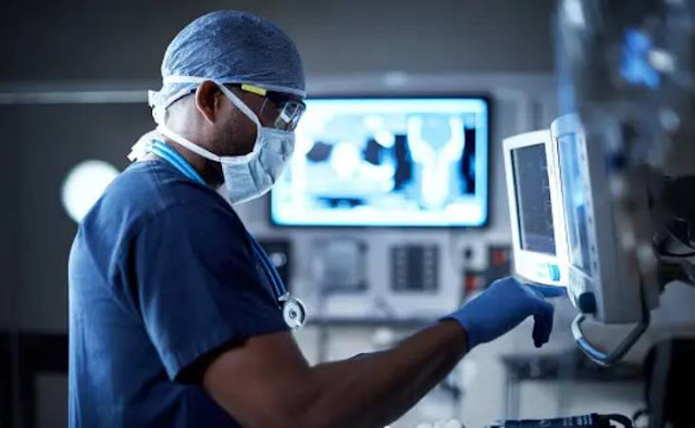
Physicians and primary care vendors regularly choose to order a selected medical imaging examination based totally on a patient’s signs and symptoms and capability diagnoses. By applying their understanding of era and human anatomy, clinical imaging technologists seize focused snap shots, enabling healthcare specialists to examine areas of a affected person’s frame for signs and symptoms of infection or sickness. Several varieties of clinical imaging technology are utilized in numerous sides of drugs.
Computed Tomography
Computed tomography, regularly known as CT or CAT scanning (Computerized Axial Tomography), is a medical imaging technology that uses X-ray radiation. Images are created when X-rays pass thru a patient’s frame and specialised detectors capture the exiting X-rays, converting this records to a visible image. Computed tomography scans take numerous photographs through non-stop sections of a patient’s frame or frame component. This creates a fixed of pass-sectional images that provide records approximately bones, tissues, and blood vessels read more:- learninfotechnologyies
Computed tomography scans may be greater powerful than undeniable X-rays because they're extra precise, however they do require better doses of radiation. Doctors regularly use CT scans to diagnose inner accidents after an coincidence, locate a tumor, or come across a ailment which include most cancers. CT scans can also assist doctors screen a affected person’s progress in improving from an damage, which include a damaged leg, or from a circumstance, which include coronary heart ailment.
Magnetic Resonance Imaging
Magnetic resonance imaging (MRI) utilizes superconducting magnets and radio waves to shape photographs in preference to ionizing radiation. An MRI system consists of a large magnet that creates a magnetic field. An MRI test makes use of a sturdy magnetic subject and radio waves to generate pix of organs and tissues. Doctors select to apply MRI when they need to investigate a patient’s ligaments and tendons, smooth tissues, or organs. MRI of the mind can assist docs diagnose strokes, tumors, eye issues, aneurysms, and different situations.
A physician can also use MRI to take a look at the scale of a affected person’s coronary heart, the consequences of a coronary heart attack, or the inflammation of blood vessels. Magnetic resonance imaging also can assist doctors hit upon tumors or most cancers in a patient’s liver, breast, ovaries, kidney, pancreas, and different organs read more:- themeisle1403
Vascular Interventional Radiography
Vascular interventional radiology permits medical doctors to treat diverse situations thru angioplasty, stenting, thrombolysis, and different minimally invasive approaches. Vascular interventional radiology can utilize more than one techniques and imaging procedures, inclusive of computed tomography, ultrasounds, and X-ray fluoroscopy.
Many vascular interventional radiology processes contain an interventional radiologist passing a needle thru a small incision within the affected person’s skin to the treatment region. Some tactics contain small catheter tubes or wires that docs can use to navigate thru a affected person’s frame. Vascular interventional radiology allows doctors deal with blood vessel disease, clear up troubles in dilated or blocked veins, guide benign tumor healing procedures, and do away with kidney or gallbladder stones.
Sonography
Sonography is a form of clinical imaging, also referred to as ultrasound imaging, that is predicated on sound waves. Ultrasound technologists place gel on the place in which they may be going to seize an photograph. A probe, called a transducer, is then positioned on or in the affected person’s body. In order to create photographs, sound waves are sent into the body and contemplated lower back into the transducer, which generates electric signals that are converted to visible pix. The method does now not contain any radiation, however produces a real-time transferring image.
Doctors regularly use sonography to test on fetal boom and development. Ultrasound examinations also can help diagnose conditions when a affected person has signs regarding ache, swelling, or infection. Doctors use ultrasounds to study a affected person’s heart, gallbladder, liver, kidneys, brain, backbone, and different organs read more:- technoid1403
- Get link
- X
- Other Apps
Popular Posts
Makeup Essentials Everyone Should Own + Popular Holy Grails
- Get link
- X
- Other Apps

